VetRepro

Stories and thoughts about small animal reproduction and ultrasound


Leave a comment

Aging of the canine prostate

Hi everybody!

In the next posts we will discuss a little bit about the prostate of the dog and will try to distinguish mythology from real life!

We all know that the prostate gland is changing in accordance with the age of the dog. These age-related changes have been documented in the veterinary literature. It is well known for example that the prostate gland commonly develops benign prostatic hyperplasia (BPH) in intact male dogs over 5 years, while in dogs older than 6 years signs suggestive of prostatic disease are commonly found.

The incidence of prostatic diseases has risen steadily over the past years as a result of dog’s life expectancy increase!  The overall median age of death is 11 years approximately and, according to the literature, there is a tendency to increase more. This is the result of several different factors, such as better management, better nutrition, owner education and improved veterinary care and prevention.

Most common prostatic diseases such as BPH, and cysts are generally asymptomatic at their onset and their early detection would allow the veterinarian to plan specific follow up and to recommend effective therapeutic protocols. So, a non-invasive screening of the prostate status and health would be advisable as a part of a preventive medicine program of geriatric diseases in dogs.

will-langenberg-11534

The physio-pathological process of aging of the prostate gland has been well studied, but still no information is available about at what age, how often and even whether a screening program of the prostate health should be recommended in dogs. To define a screening program, the age when the examination should begin, is the first decision to be made. Due to different breed’s expected longevity, a dog of a certain age might be considered as geriatric in large breeds, and not geriatric in small breeds. For instance, small-breed dogs become geriatric at about 11 years, whereas giant-breed dogs at 7 years. Longevity in crossbred dogs exceeds that of purebred dogs by 1.2 years and increasing bodyweight is negatively correlated with life expectancy. Thus, the age for the early detection of abnormalities in the prostate could vary in dogs of different breeds…

On the basis of all these, our group decided to perform a study in order to estimate the recommended age for a preventive ultrasonographic examination of the prostate in the dog. In the forthcoming posts, we will present you the design of our study! So stay tuned!

Till then, enjoy your life and love your pets!


1 Comment

Small animals’ genital tract: Ultrasound vs microscope

On Sunday 18th of September a day conference “Small animals’ genital tract: Ultrasound vs microscope” was organized by Dr. Constantinos Teliousis  (good old friend) and me at the Hellenic Pasteur Institute in Athens.

More than 120 veterinarians attended the event! And they were satisfied!

All the aspects of ultrasound examination of the male and female genital tract of the dog and cat were covered , by answering the following three questions:
-When to perform ultrasound of the genital tract?
-Why to perform ultrasound?
-And finally how to perform ultrasound?

Normal and pathological sonographic appearance of the prostate, the testicles, the scrotum, the uterus and the ovaries of the dog and cat were presented, with several pictures and videos.

Pathology of the genital tract and the mammary glands of dog and cat was thoroughly covered by Constantinos. Cytological and histological findings of their most common diseases were also briefly reviewed. Constantinos also presented several useful “tips and tricks” for the clinical small animal practitioner!

Finally, a special thanks to our sponsor PetLine for funding and for helping organizing the whole thing!


1 Comment

Pregnancy diagnosis in the bitch and queen

Today I will try to summarize different methods that can be used for pregnancy diagnosis in the bitch and queen.

First of all, we have to remember that the embryo enters uterus on day 5-8. Attachment and maternal recognition occur on days 13-15 in the bitch and days 12-13 in the queen.

Clinical signs
The clinical signs of pregnancy are variable and unreliable. The primiparous bitch might show slight enlargement of teats on days 35 – 45.

Palpation
Abdominal palpation may give the veterinarian several diagnostic information. Segmental dilations can be palpated in the bitch from day 24 – 35 after ovulation and in the cat between day 16 – 26.
On the 3rd week the conceptuses are 15mm, round in shape, firm, well separated from each other. On week 4 they are 25mm, more oval, and on week 5:30-35mm, oval, soft, no separate structures anymore. After day 35 fetal vesicles enlarge and palpation is less accurate. Later on, fetal heads and bodies might be palpated. In large breeds and in bitches with tense abdomen or small litters palpation may be false negative.
Estimation of litter size is not possible. Finally, be careful: palpation needs to be as gentle as possible!

Ultrasound
Ultrasonography is the best method, safe and accurate. It allows the assessment of pregnancy status, and viability of the fetuses.
Gestational sacs are visible as early as day 18-20 after LH peak, but its recommend to examine not before day 21-25 because the small fluid-filled structures may be obscured by intestinal gas earlier.
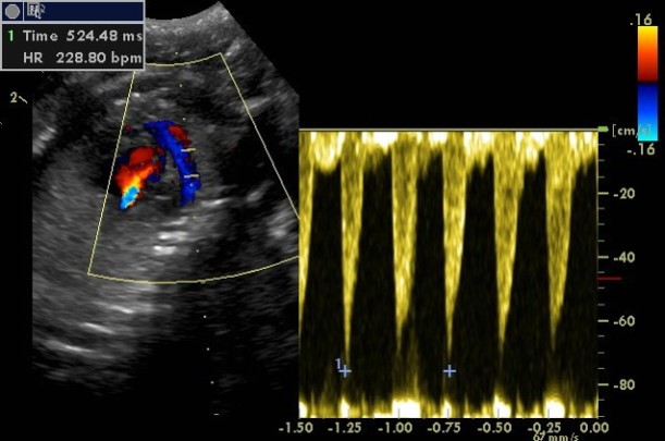
Fetal heartbeat can be detected from days 16-25 in the queen and from days 23-28 in the bitch. Fetal heart rate should be more than 200 beats per minute as decreasing rate indicates fetal stress. Several ultrasound methods can be used to calculate it.

hr

Estimation of fetal heart rate with Pulsed Wave Doppler

After day 28 in the queen and days 34-36 in the bitch fetal movements are present.
Parturition time can be estimated with measurement of gestational sac diameter, crown-rump length and head diameter.
In contrary with what is believed, ultrasonography is NOT the method of choice for assessment of litter size. Counting number of fetuses is difficult, especially in large litters.

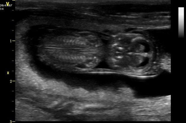
d39

Canine fetus, day 39

Radiography
Enlarged fluid filled uterine horns can be observed by day 21-42 but they cannot be distinguished from that seen with uterine disease, pyometra for example.
First skeletal mineralization occurs by day 42. The degree of fetal mineralization can be used to assess gestational age and predict whelping date.
Radiography is useful more in late pregnancy, when it can be used to identify litter size and fetal size in relation to birth canal.
During the first trimester of pregnancy (organogenesis) there is high risk of radiation damage, but later in gestation this risk is minimal and not greater for fetuses than for the bitch.
Signs of fetal death after 24 hours include presence of gas within or around the fetus and collapse of the axial skeleton.

Hormones
– Relaxin
Is produced by placenta. A serum test is available and can be used from day 24 after ovulation. Estimation of relaxin gives no information about number or viability of the fetuses. The test remains positive for an undetermined time after pregnancy loss occurs. In general, it is not commonly used.
– Progesterone
Progesterone is not indicative! It remains high in all normal dogs during diestrus regardless of breeding status or pregnancy as in the bitch is produced exclusively from corpus luteum. Some studies have documented slight difference in the pattern of progesterone of pregnant and not pregnant bitches but interpretation needs several measurements of serum concentration.
– Prolactin, FSH and estrogen
There are differences in concentrations but not practical to use.

Several other things about pregnancy diagnosis can be written and further analyzed, so if you want we can always come back later!

Thanks for reading! See you soon!