VetRepro

Stories and thoughts about small animal reproduction and ultrasound


Leave a comment

Extreme Breeding

EVSSAR, as the leading veterinary society on small animal reproduction and as an associate member of FECAVA, salutes WSAVA‘s endorsement on FVE/FECAVA Position Paper on Healthy Breeding!

https://www.fecava.org/en/newsroom/news/news-cat/wsava-endorses-fvefecava-position-paper-on-healthy-breeding.htm

Here you can read and download the FECAVA, FVE Position Paper on breeding healthy dogs.

Just to remember, the discussion of “Ethics in Animal Breeding” has began in 2017, at Vienna, from Prof. W. Farstad! Some very interesting lectures were also given by excellent scientists and experts at Venice, during EVSSAR’s 21st Congress:

-Ethical considerations in small animal reproduction, by H. Ovregaard.
-How do repro experts deal with ethical issues- a survey, by S. Arlt.
-Antibiotics for bacterial infections in veterinary medicine – sustainable use secures future health, by H. SØrum.

All these are nothing more but an excellent starting point.  We promise that we, EVSSAR, will continue to discuss Ethics in our future congresses!

Stay tuned!

Featured image by JC Gellidon on Unsplash


Leave a comment

Aging of the canine prostate

Hi everybody!

In the next posts we will discuss a little bit about the prostate of the dog and will try to distinguish mythology from real life!

We all know that the prostate gland is changing in accordance with the age of the dog. These age-related changes have been documented in the veterinary literature. It is well known for example that the prostate gland commonly develops benign prostatic hyperplasia (BPH) in intact male dogs over 5 years, while in dogs older than 6 years signs suggestive of prostatic disease are commonly found.

The incidence of prostatic diseases has risen steadily over the past years as a result of dog’s life expectancy increase!  The overall median age of death is 11 years approximately and, according to the literature, there is a tendency to increase more. This is the result of several different factors, such as better management, better nutrition, owner education and improved veterinary care and prevention.

Most common prostatic diseases such as BPH, and cysts are generally asymptomatic at their onset and their early detection would allow the veterinarian to plan specific follow up and to recommend effective therapeutic protocols. So, a non-invasive screening of the prostate status and health would be advisable as a part of a preventive medicine program of geriatric diseases in dogs.

will-langenberg-11534

The physio-pathological process of aging of the prostate gland has been well studied, but still no information is available about at what age, how often and even whether a screening program of the prostate health should be recommended in dogs. To define a screening program, the age when the examination should begin, is the first decision to be made. Due to different breed’s expected longevity, a dog of a certain age might be considered as geriatric in large breeds, and not geriatric in small breeds. For instance, small-breed dogs become geriatric at about 11 years, whereas giant-breed dogs at 7 years. Longevity in crossbred dogs exceeds that of purebred dogs by 1.2 years and increasing bodyweight is negatively correlated with life expectancy. Thus, the age for the early detection of abnormalities in the prostate could vary in dogs of different breeds…

On the basis of all these, our group decided to perform a study in order to estimate the recommended age for a preventive ultrasonographic examination of the prostate in the dog. In the forthcoming posts, we will present you the design of our study! So stay tuned!

Till then, enjoy your life and love your pets!


Leave a comment

Ethics and small animal reproduction

Helen, a good friend of mine, a vet from Norway, showed me a couple of months ago a video that was viral in Norway. A video trying to inform and “sensitize” owners about the possible problems that brachycephalic breeds of dogs have. You can watch it by just clicking the following link:

https://www.youtube.com/watch?v=oghf4Figs_Q

Indeed, certain breeds of dogs and cats are prone to difficult, obstructive breathing, because of the shape of their head, muzzle and throat. This pathological syndrome is known as “brachycephalic syndrome” and can lead to severe respiratory distress.  According to ACVS (https://www.acvs.org/small-animal/brachycephalic-syndrome), “the term Brachycephalic Syndrome refers to the combination of elongated soft palate, stenotic nares, and everted laryngeal saccules.

So one obvious question someone could have is:

  • Should breeders reproduce animals of brachycephalic breeds the same way they have been doing till today? 

And not only that! In our modern “fast” world, several questions, concerning Ethics and small animal reproduction, arise:

  • Should we spay or not dogs and cats?
  • Which is the most suitable age to spay a dog or a cat?
  • Why retained testicles are so common in dogs and cats?
  • Which way can we avoid inherited diseases?

And further more:

  • Should we choose sex in fetuses?
  • What about cloning of small animals?
  • Cryopreservation of small animal embryos?
  • etc etc etc

And finally, who is leading and who should lead research on reproduction? Big pet-food companies? Pharmaceutical companies? Maybe breeders? Our clients? And is there any place for Ethics? What is the role of Education?

Hot questions! Impossible to answer all of them! But here are the good news: In the forthcoming 20th International Congress of Small Animal Reproduction that will be held in Vienna (Austria) from June 29th to July 1st 2017, the opening speech is exactly what we are talking about: “Ethics in Animal Breeding”, by Prof. W. Farstad! An excellent professor, vet, and breeder as well! This congress is organized by the European Veterinary Society of Small Animal Reproduction (EVSSAR), the largest society for small animal reproduction in Europe (and maybe in the world). EVSSAR aims to support continuing education of veterinarians and to promote research. More details about EVSSAR can be found here: http://www.evssar.org. The scientific program of the congress, in case you are interested, can be found here: http://www.evssar.org/wp-content/uploads/2017/04/20th-EVSSAR-program.pdf.

So at last, something seems to move… I will be back on the question above soon!

Oh I forgot: HAPPY EASTER!!!


Leave a comment

Survey for Vets!

Dear friends,
During the EVSSAR conference (Vienna, June 29th – July 1st 2017) we will organize an interactive project for our routines, experiences and opinions about ovulation timing in breeding bitches. More details can be found on EVSSAR‘s website:

www.evssar.org

For that purpose our colleague Sebastian Arlt has developed an online survey that it should not take you more than 3-4 minutes to complete. So if you are a Vet interested in small animal reproduction, click the following link (or copy it to your browser):

https://docs.google.com/forms/d/e/1FAIpQLSfMCNXwlancuEIKwKhJZEN648637jECyWrngUSrZe0OQE1xKw/viewform?usp=sf_link

Thank you in advance for your participation! Hope to see you in Vienna,
On behalf of Sebastian Arlt and the EVSSAR Board,
George


1 Comment

Small animals’ genital tract: Ultrasound vs microscope

On Sunday 18th of September a day conference “Small animals’ genital tract: Ultrasound vs microscope” was organized by Dr. Constantinos Teliousis  (good old friend) and me at the Hellenic Pasteur Institute in Athens.

More than 120 veterinarians attended the event! And they were satisfied!

All the aspects of ultrasound examination of the male and female genital tract of the dog and cat were covered , by answering the following three questions:
-When to perform ultrasound of the genital tract?
-Why to perform ultrasound?
-And finally how to perform ultrasound?

Normal and pathological sonographic appearance of the prostate, the testicles, the scrotum, the uterus and the ovaries of the dog and cat were presented, with several pictures and videos.

Pathology of the genital tract and the mammary glands of dog and cat was thoroughly covered by Constantinos. Cytological and histological findings of their most common diseases were also briefly reviewed. Constantinos also presented several useful “tips and tricks” for the clinical small animal practitioner!

Finally, a special thanks to our sponsor PetLine for funding and for helping organizing the whole thing!


Leave a comment

Proceedings of the ISCFR-EVSSAR Congress – Paris, 2016

The abstracts of the 8th International Symposium on Canine and Feline Reproduction (ISCFR-EVSSAR, Paris 2016) are available on line at IVIS website! If you want to download them, click here! Remember, you have to be registered to IVIS in order to have full access…

This excellent Symposium was the result of skillful and proficient work of the ISCFR International Organizing Committee members Prof. Michelle Kutzler, Prof. Pierre Comizzoli, Prof. Gary England and Prof. John Verstegen, in collaboration with the Scientific Program committee, and the Local Organizing Committee chaired by Prof. Alain Fontbonne and Associate Prof. Karine Reynaud. Of course we have to mention the excellent work of  all the members of the EVSSAR board!

Finally a big big thank you to all the vets from all over the world that participated actively and presented the results of their latest scientific work on all the fields of small animal (and not only) reproduction!

PS: The party organized from the local organizing committee was just awesome!  Thank you guys!

dsc06337-small


2 Comments

News from the ISCFR-EVSSAR Congress – Paris, 2016

The 8th Quadrennial International Symposium on Canine and Feline Reproduction which is a  Joint Meeting with the 19th EVSSAR Congress, just finished. It was perfectly organized in Paris, the city of light, from 22 to 25 of June.

The scientific level of all speakers was very high and the program covered all the aspects of small animal reproduction science.

The results of our latest research projects were successfully presented:

– Ultrasound elastography of the normal canine prostate and testicles (G. Mantziaras, G.C. Luvoni).

-First documented report of CHV-1 infection of a pregnant bitch in Greece.  (G. Mantziaras, K. Teliousis, O. Mavropoulou, A. Pseftogas, G.C. Luvoni).

It was also a great chance to meet good friends from all over the word, discuss with them, and have a nice party on Friday night!

Finally, I was one of the three new EVSSAR’s Board members that have been elected during the General Assembly! A big big thank you to all those that honored me with their vote! I promise to do my best and work hard the following years!!!

We’ll be back soon and till than,

Take care of yourselves!


1 Comment

Pregnancy diagnosis in the bitch and queen

Today I will try to summarize different methods that can be used for pregnancy diagnosis in the bitch and queen.

First of all, we have to remember that the embryo enters uterus on day 5-8. Attachment and maternal recognition occur on days 13-15 in the bitch and days 12-13 in the queen.

Clinical signs
The clinical signs of pregnancy are variable and unreliable. The primiparous bitch might show slight enlargement of teats on days 35 – 45.

Palpation
Abdominal palpation may give the veterinarian several diagnostic information. Segmental dilations can be palpated in the bitch from day 24 – 35 after ovulation and in the cat between day 16 – 26.
On the 3rd week the conceptuses are 15mm, round in shape, firm, well separated from each other. On week 4 they are 25mm, more oval, and on week 5:30-35mm, oval, soft, no separate structures anymore. After day 35 fetal vesicles enlarge and palpation is less accurate. Later on, fetal heads and bodies might be palpated. In large breeds and in bitches with tense abdomen or small litters palpation may be false negative.
Estimation of litter size is not possible. Finally, be careful: palpation needs to be as gentle as possible!

Ultrasound
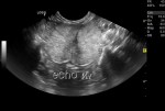
Ultrasonography is the best method, safe and accurate. It allows the assessment of pregnancy status, and viability of the fetuses.
Gestational sacs are visible as early as day 18-20 after LH peak, but its recommend to examine not before day 21-25 because the small fluid-filled structures may be obscured by intestinal gas earlier.
Fetal heartbeat can be detected from days 16-25 in the queen and from days 23-28 in the bitch. Fetal heart rate should be more than 200 beats per minute as decreasing rate indicates fetal stress. Several ultrasound methods can be used to calculate it.

hr

Estimation of fetal heart rate with Pulsed Wave Doppler

After day 28 in the queen and days 34-36 in the bitch fetal movements are present.
Parturition time can be estimated with measurement of gestational sac diameter, crown-rump length and head diameter.
In contrary with what is believed, ultrasonography is NOT the method of choice for assessment of litter size. Counting number of fetuses is difficult, especially in large litters.

d39

Canine fetus, day 39

Radiography
Enlarged fluid filled uterine horns can be observed by day 21-42 but they cannot be distinguished from that seen with uterine disease, pyometra for example.
First skeletal mineralization occurs by day 42. The degree of fetal mineralization can be used to assess gestational age and predict whelping date.
Radiography is useful more in late pregnancy, when it can be used to identify litter size and fetal size in relation to birth canal.
During the first trimester of pregnancy (organogenesis) there is high risk of radiation damage, but later in gestation this risk is minimal and not greater for fetuses than for the bitch.
Signs of fetal death after 24 hours include presence of gas within or around the fetus and collapse of the axial skeleton.

Hormones
– Relaxin
Is produced by placenta. A serum test is available and can be used from day 24 after ovulation. Estimation of relaxin gives no information about number or viability of the fetuses. The test remains positive for an undetermined time after pregnancy loss occurs. In general, it is not commonly used.
– Progesterone
Progesterone is not indicative! It remains high in all normal dogs during diestrus regardless of breeding status or pregnancy as in the bitch is produced exclusively from corpus luteum. Some studies have documented slight difference in the pattern of progesterone of pregnant and not pregnant bitches but interpretation needs several measurements of serum concentration.
– Prolactin, FSH and estrogen
There are differences in concentrations but not practical to use.

Several other things about pregnancy diagnosis can be written and further analyzed, so if you want we can always come back later!

Thanks for reading! See you soon!


Leave a comment

The normal parturition of the dog

So at last, time has come! Your dog is ready to be a mother and you are ready to become grandmas and grandpas!

At the following lines we will try to explain you in a simplified way the physiology of canine parturition, and to present you some tips in order to be ready to welcome the new members of your family! Greek speaking people feel free to click the following link:

Ο φυσιολογικός τοκετός στη σκύλα

The first question that usually arises is “when”?

In general canine gestation (pregnancy) lasts 63 days, but can range from 59-68 days. Record the dates of all successful breedings on a calendar. For bitches bred over more than once, choose the middle date to count from. 56-57 days after breeding, start monitoring your bitch for other signs of labor. If your bitch has not whelped by 65 days, an X-ray or (better) ultrasound is recommended. Milk production begins 1-3 days prior to parturition, but this can range a lot (some bitches can produce milk 7 days before whelping while some others willnot have milk evident until after they are in labor). In primiparous lactation begins with parturition. In pluriparous lactation may begin days before parturition. The temperature of almost all bitches will drop approximately 24hours before parturition. Owners should start measure bitch’s temperature two to three times per day from day 57 of pregnancy. Most bitches will refuse to eat 4 to 24 hours before they go into labor. This is normal and expected. Do not try to force your dog to eat! If anorexia continues and does not correspond with other signs of impending parturition for more than 48, you’d better contact your vet.

Ok, now get ready for the big day! 

When delivery time is coming, you will have to prepare a few items, such as:

  • A whelping box
  • Bedding, newspapers
  • Plastic bags
  • Disinfectant and cleaning material
  • Suitable bags for disposal of placentas or stillborns (in cases that histological examination is needed)
  • Therometer
  • Pen and a table to write down the rectal temperature
  • Clock
  • Table to record timing of contractions, times of delivery
  • Gloves
  • Lubricant
  • Cotton
  • Dental floss to tie the umbilical cords
  • Scissors
  • Blankets
  • Cleaning towels
  • Marker pens to mark the puppies
  • Bottles for feeding or syringes
  • Commercially available colostrum
  • Water for the mother during labor

medium

And what happens during parturition?

The labor in dogs has three stages. The first stage (about 12h) is clinically unapparent. The bitch is restless, indifferent to food and shows nesting behavior. Uterine contractions increase and the cervix dilates. During the first uterine contractions bitches frequently change their position but stay recumbent during straining efforts. The uterine contractions are very weak and are therefore notvisible externally.

Duiring the second stage contractions will become more and more frequent. The cervix will be fully open and she will be ready for delivery. The dog needs to be calm and you need to provide a warm and quiet room for the delivery. The onset of the 2nd stage may be difficult to recognize due to the bitches movements when abdominal straining begins. The allantochorionic membrane of the first fetus appears and reaches the size of a golf ball. The bitch breaks it by licking. The delivery of the head needs the greatest effort, the rest usually follows without delay. The expulsion of the first puppy may last one hour. About 40% of puppies are expulsed in posterior presentation, which is absolutely normal. The puppies remain attached to the umbilical cord up to its rupture post partum. After expulsion of the first puppy the bitch rests for some time and licks the puppy until it shows signs of viability. She licks the vulvar discharge and eats the placenta which is expelled within 10-15 minutes (this is the third stage). Sometimes, more pups may be delivered before the membranes are expelled. A delay up to 24 hours may occur until the expulsion of all fetal membranes is finished. The bitch may try to eat the fetal membranes. Because this may lead to vomiting and/or diarrhoea, it is recommended that the membranes are removed. The second and third stages are then repeated. After 30-120 minutes delay,  straining recommences. The interval between two puppies is about 30-60 minutes. The second stage  lasts 3-12 hours in bitches.

Stay tuned and till then, congratulations on your new arrivals!!!